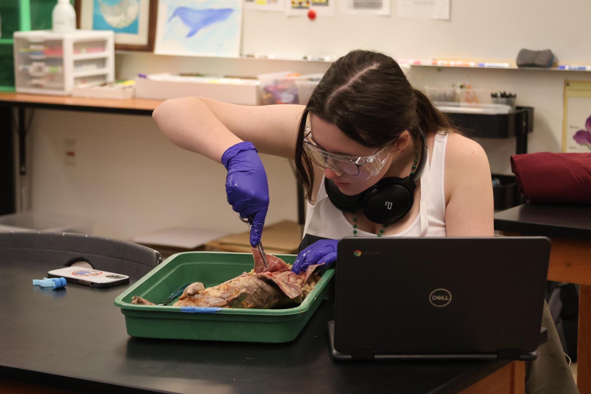
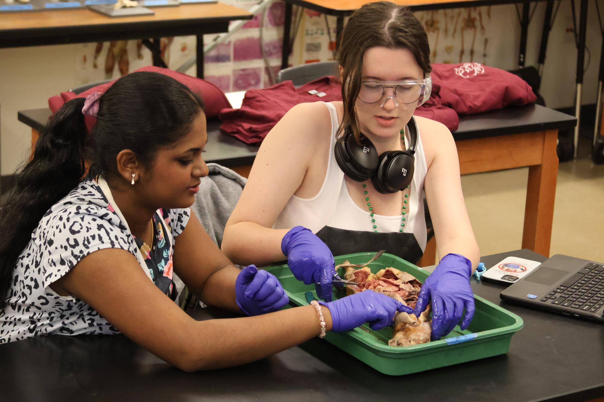
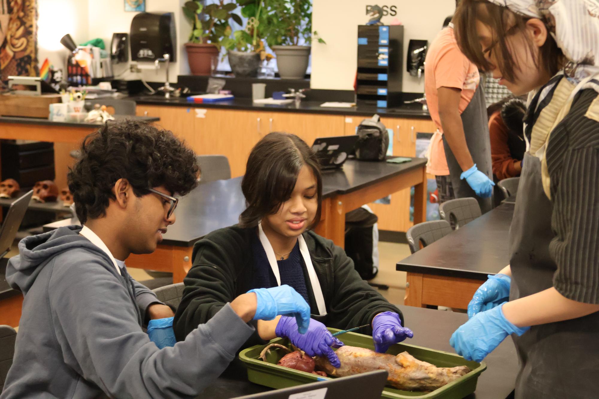
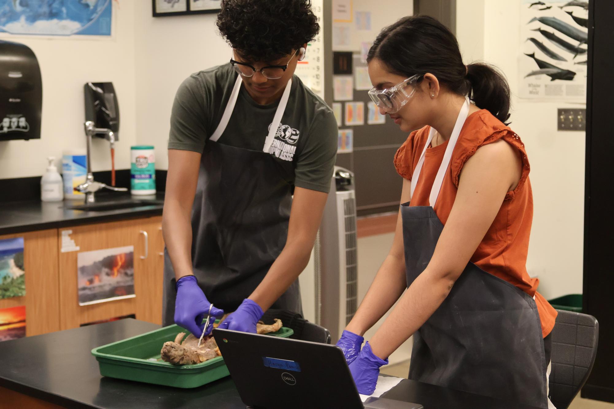
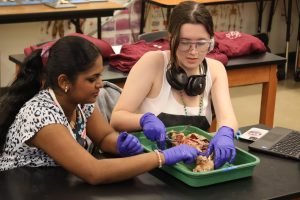
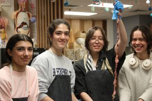
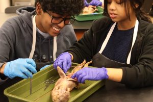
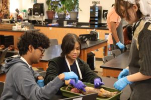
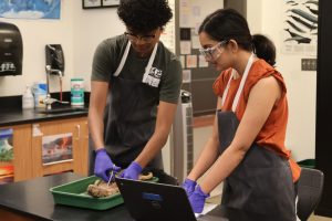
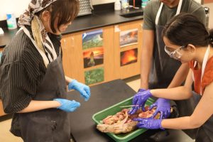
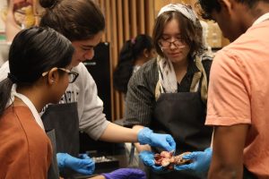
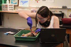
For their last meeting of the year, Anatomy & Dissection Club held a mink dissection in Mr. Eric Scheiber’s room after school on Thursday, March 21. The mink was the largest and most complex specimen club members explored so far. However, due to its compactness, the club completed the dissection in one sitting, unlike the two-part fetal pig study last year.
“Last year, we did fetal pigs, so we wanted to do something similar for our big dissection this year, and minks are some of the more common big ones,” Anatomy & Dissection Club President Sanjay Balasubramanian ‘24 said.
The minks’ veins and capillaries were not dyed before they were preserved, vacuum-sealed, and shipped to Westwood, complicating club members’ efforts to identify the animal’s mammalian internal organs. However, to make the dissection easier, the specimens were mostly de-furred, except for their ears and the tips of their tails.
“It’s a lot of work [to dissect a furry specimen] because the furs get messed up so if you make a cut,” Anatomy & Dissection Club Vice President Nicole Wang ‘24 said. “It’s like cutting a really lush rug. It’s kind of difficult to part [the fur] and see [the insides] because all of the hairs are going in different directions, and you can’t see your scalpel.”
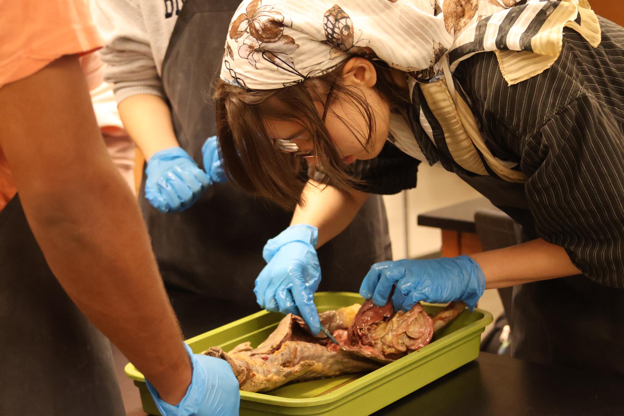
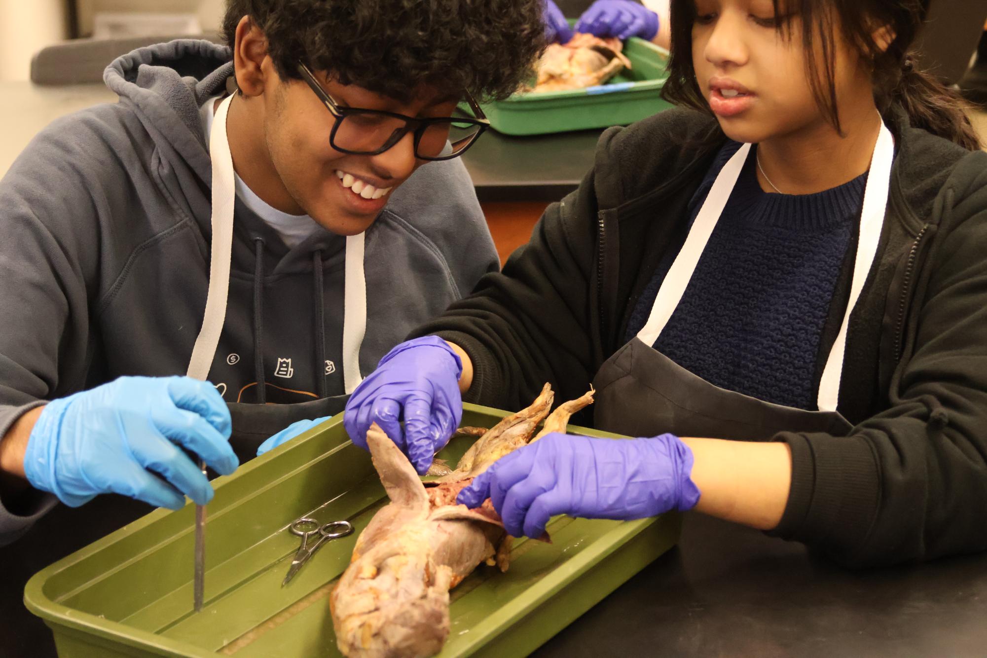
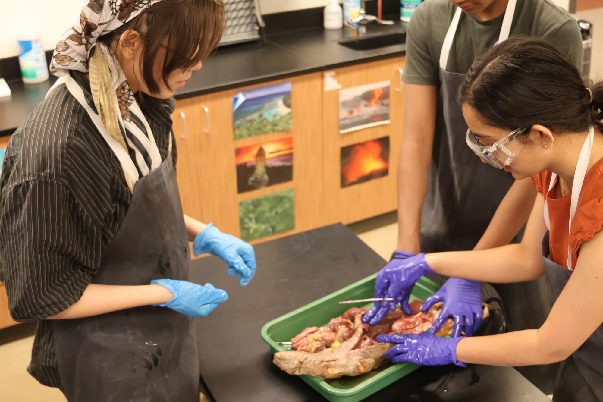
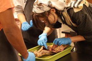
Prior to the dissection, club members examined the mink’s external features, including its small rounded ears and elongated head. Then, using scissors, they carefully made a midline incision in the mink’s abdominal and thoracic cavities. Finally, they peeled back the skin to expose the internal structures, identifying and removing the specimen’s organs.
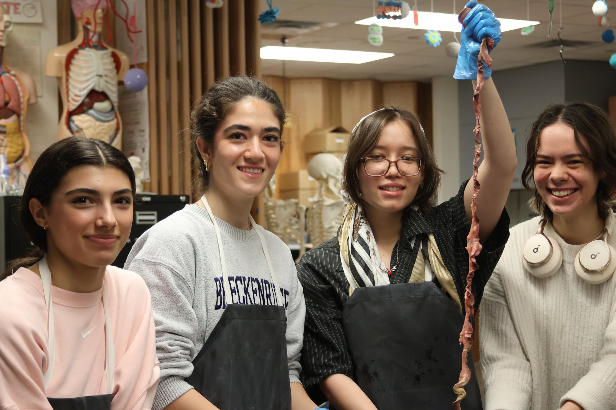
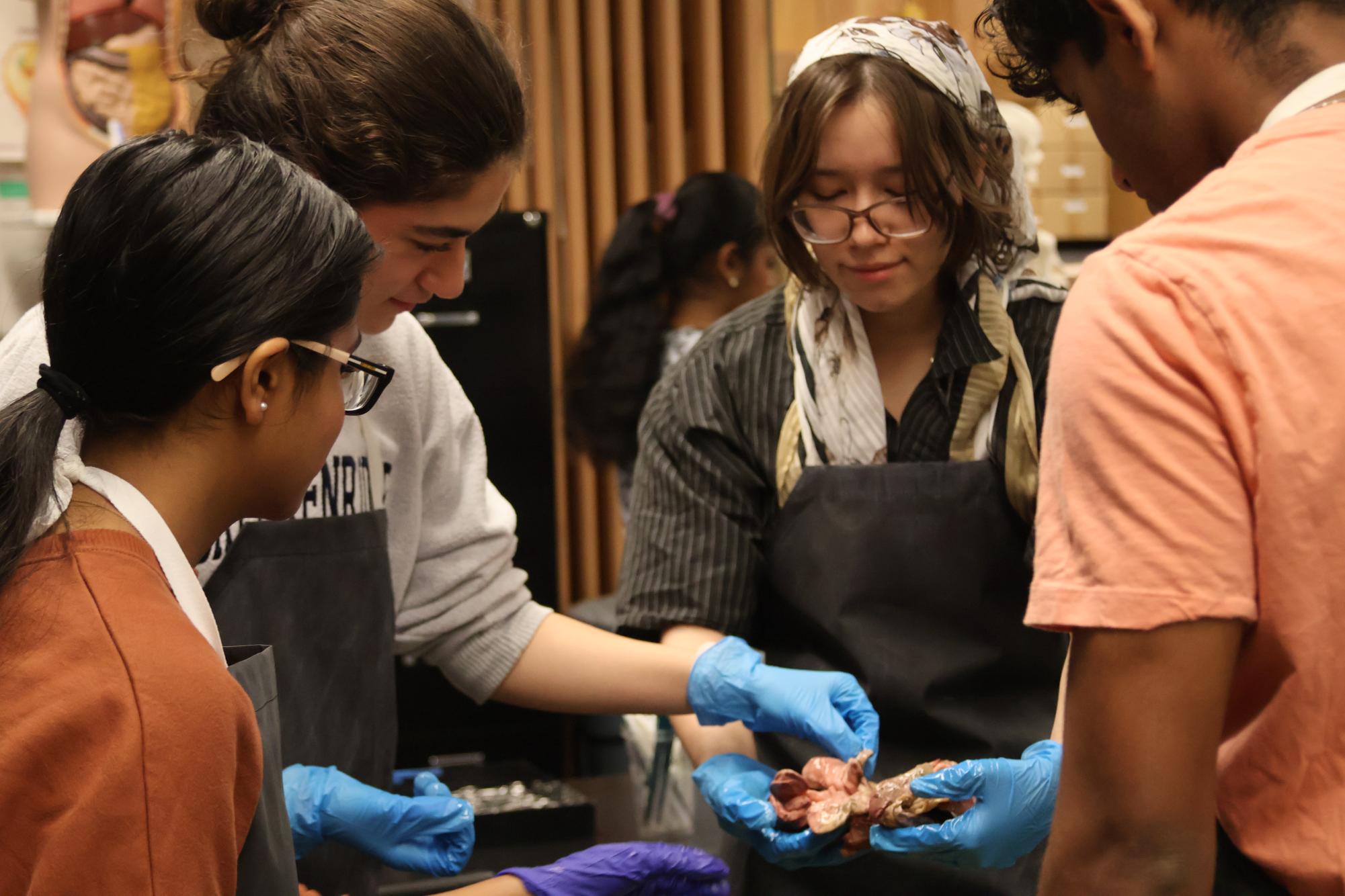
“We pulled out the intestines, then we took out specific organs and examined those,” club member Rahul Suresh ‘25 said. “I found the intestines [to be] the most interesting because they were surprisingly long for how small of a creature it was. It was a different kind of specimen than the smaller specimens we’ve done before so it was pretty cool.”
Club members collaborated to work through some challenges, including cutting through the ribcage and taking off the tail, which was skeletally connected to the spinal cord. Additionally, some groups faced unforeseen issues.
“I squeezed the intestine too hard and it squirted,” club member Preethi Ram ‘25 said. “It tasted like chemicals.”
Due to its self-paced nature, the dissection taught members about the mink’s organ systems, such as its cardiovascular system, which was small, considering the animal’s relatively large size. These discoveries allowed medical students to apply what they learned in class and prepare for more real-life experiences after high school.
“We’ve been part of the Anatomy & Dissection Club for around two years now and we’re aspiring health students,” Suresh said. “We want to be doctors, so doing dissections and getting experience is really captivating for us.”
According to Wang, the dissections were tiring to prepare, but ultimately worth it. Throughout the year, they increased in complexity to build up members’ skills in preparation for the minks, including some call-backs from previous years. Additionally, the club has been hosting increasingly advanced dissections every year, and will likely feature more complex organisms next year as well.
“I think [this year] was great,” Balasubramanian said. “We were very well organized. We had a lot of good dissections as well. I did the squid sophomore year and it was really cool, coming back to it, because it’s not common for dissections. This year, we did a couple of new dissections and we did bigger organisms. We learned a lot about anatomy. Familiarizing yourself with the anatomy of these creatures can help you understand their functions in future biology classes.”


![Holding her plate, Luciana Lleverino '26 steadies her food as Sahana Sakthivelmoorthy '26 helps pour cheetos into Lleverino's plate. Lleverino was elected incoming Webmaster and Sakthivelmoorthy rose to the President position. "[Bailey and Sahiti] do so much work that we don’t even know behind the scenes," Sakthivelmoorthy said. "There’s just so much work that goes into being president that I didn’t know about, so I got to learn those hacks and tricks."](https://westwoodhorizon.com/wp-content/uploads/2025/05/IMG_0063-1200x1049.jpg)


