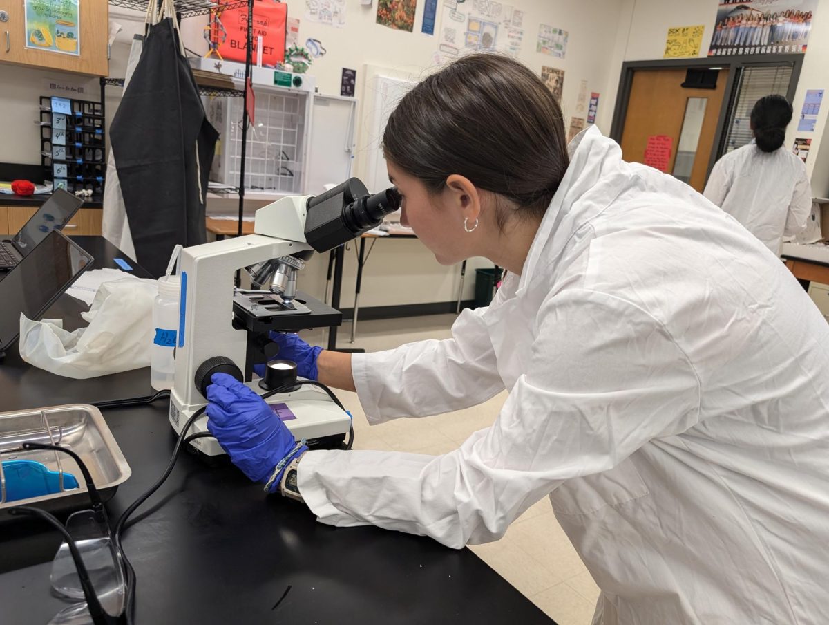
Diving deep into their microbiomes unit, Ms. Avni Kantawala’s Medical Microbiology class participated in a Simple Stain Lab on Monday, Sept. 23, where they used fluorescent staining to examine the cells from their cheek.
“We basically took a sample of our oral cells by swabbing the inside of our cheek,” Ninju Prem ‘25 said. “Then we stained it with [a dye called] Methylene blue, and looked at the cells under the microscope.”
The lab offered students the opportunity to learn more about the procedures of how to stain cells. This process involves using a permanent, dark blue pigment dye to color the cell samples, which in turn allows for the cells to be more visible under the lens of the microscope.
“[My favorite part of the lab was when we] dyed the cell sample,” Cole Osborn ’25 said. “It’s cool seeing how [the color] translates to the cells that are on [the microscope slide].”
After the dye set, students began to observe the unique details of their own cheek cells by viewing the contents on the microscope slide.
“[The cells] were shaped like a butterfly,” Angana Dahal ‘25 said. “[There were] a lot of blue dots where I could tell the DNA was.”
By looking at the cells in their own bodies, students were able to connect their learning about their microbiome to real life. Many students expressed their interest in the lab, enjoying various aspects of it, from dyeing the cells to viewing them under the microscope. This lab was in preparation for a future lab where they will stain bacteria, which are much smaller than cells.
“I like microscopes [and] so I [especially] like looking at cells under the microscope,” Prem said. “I thought that was really cool.”


![Holding her plate, Luciana Lleverino '26 steadies her food as Sahana Sakthivelmoorthy '26 helps pour cheetos into Lleverino's plate. Lleverino was elected incoming Webmaster and Sakthivelmoorthy rose to the President position. "[Bailey and Sahiti] do so much work that we don’t even know behind the scenes," Sakthivelmoorthy said. "There’s just so much work that goes into being president that I didn’t know about, so I got to learn those hacks and tricks."](https://westwoodhorizon.com/wp-content/uploads/2025/05/IMG_0063-1200x1049.jpg)

