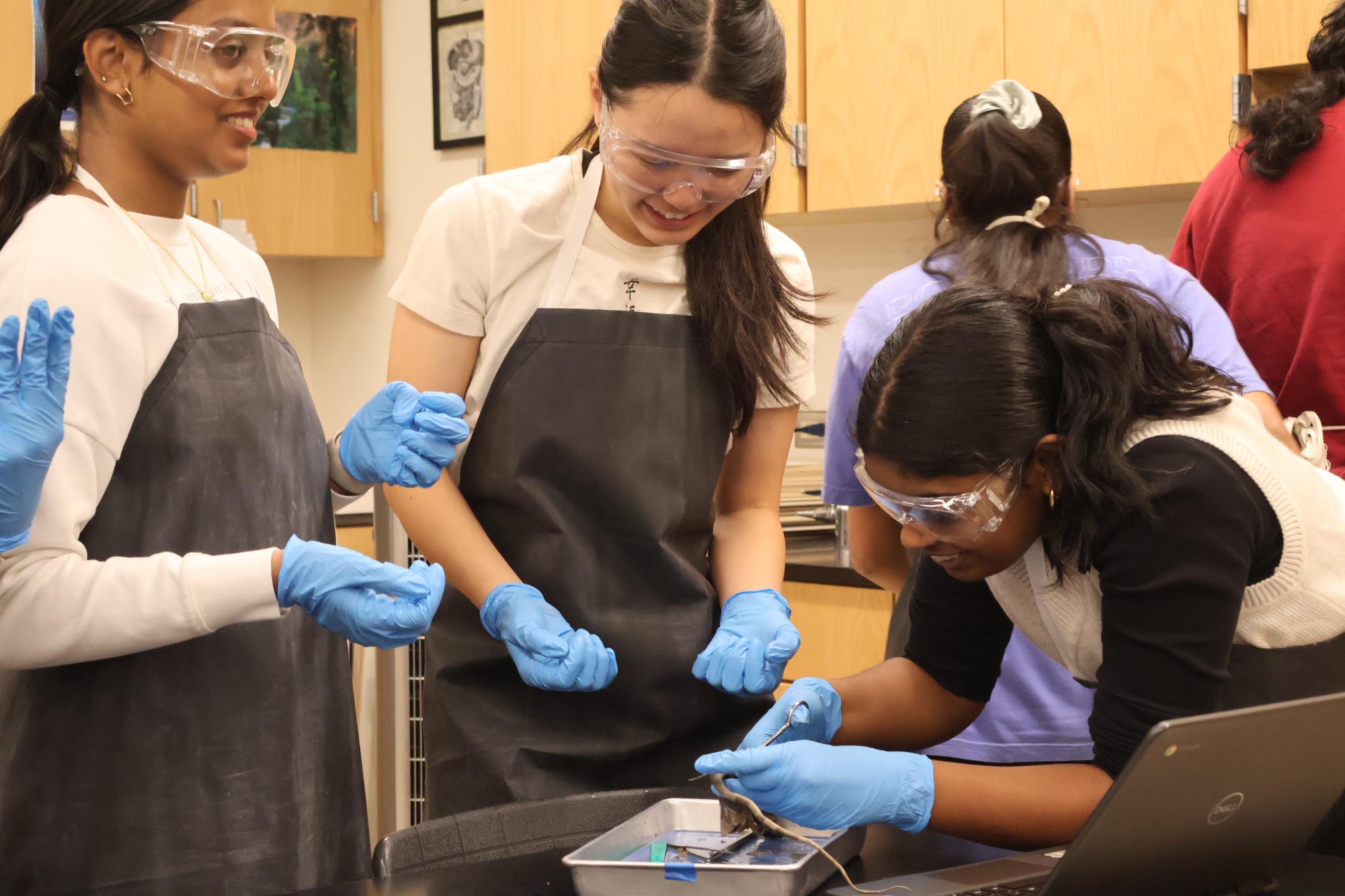
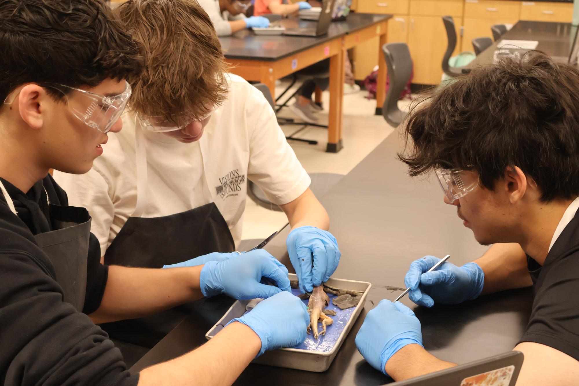
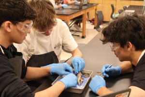
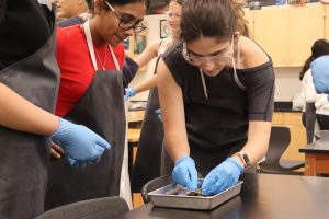
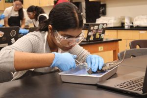
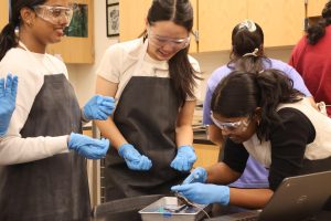
Amidst bright lights and the thick scent of formaldehyde, the Anatomy & Dissection Club dissected iguanas in Mr. Eric Scheiber’s room on Friday, March 28. In line with the club officers’ decision to introduce multiple new specimens to the club this year, this dissection allowed students practice with reptiles, while featuring an organism that the club has never examined before.
“It’s not often that we do a reptilian specimen, so this was the first time I’ve ever participated in an iguana dissection,” Mr. Scheiber said. “It’s a reptile that we can find on land and in aquatic environments, so it’s got diverse behaviors and a variety of adaptations, and it was good exposure for students.”
Although the iguana specimens that arrived were all juveniles, and therefore not as large as the club officers expected, they provided a useful stepping stone between the easier dissections and more complex dissections this school year.
“The idea was to have [club members] practice [earlier in the year], because pigeons are relatively easy to dissect. [For] iguanas, [it is] harder to get to the little nooks and crannies because of their leathery skin, so we thought [it would be] newer and just bigger and more exciting,” Anatomy & Dissection Club President Mishree Narasaiah ‘25 said. “A&D’s never done an iguana before, and I feel like most people haven’t experienced an iguana dissection either.”
Following an instructional video, club members learned about the iguana’s anatomical adaptations, such as its shedding habits and the effect of death on decreased blood flow and subsequent darkening of scale patterns. The video also featured a timelapse display of beetles eating the remaining iguana flesh to reveal the intricate skeletal structure underneath.
“Some unique features we [observed] definitely [included] the tongue,” Narasaiah said. “It was different because it was pretty small but it was so thick, and you can see how [iguanas] use it to catch prey. And then you see the scales on their necks and backs, and when you feel them, it’s also pretty nice. And, of course, they use [their tails] for balancing and everything. We thought that was really cool because they were stuck in a rigid position but still super structurally sound.”
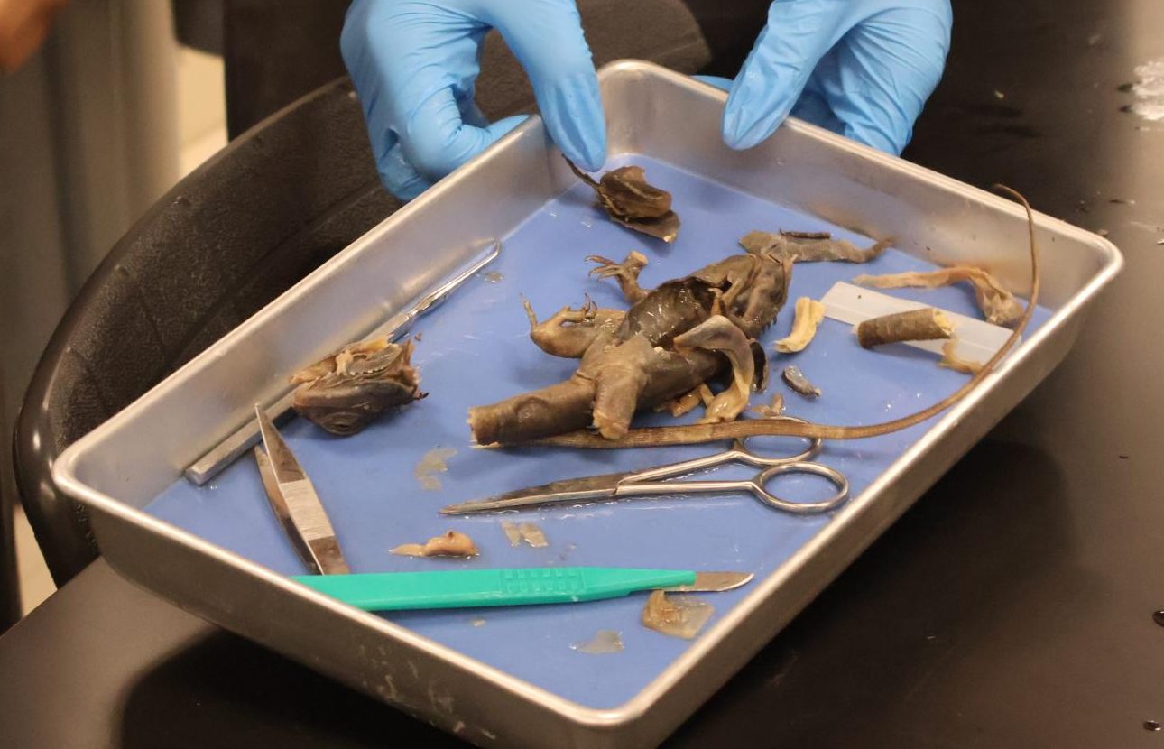
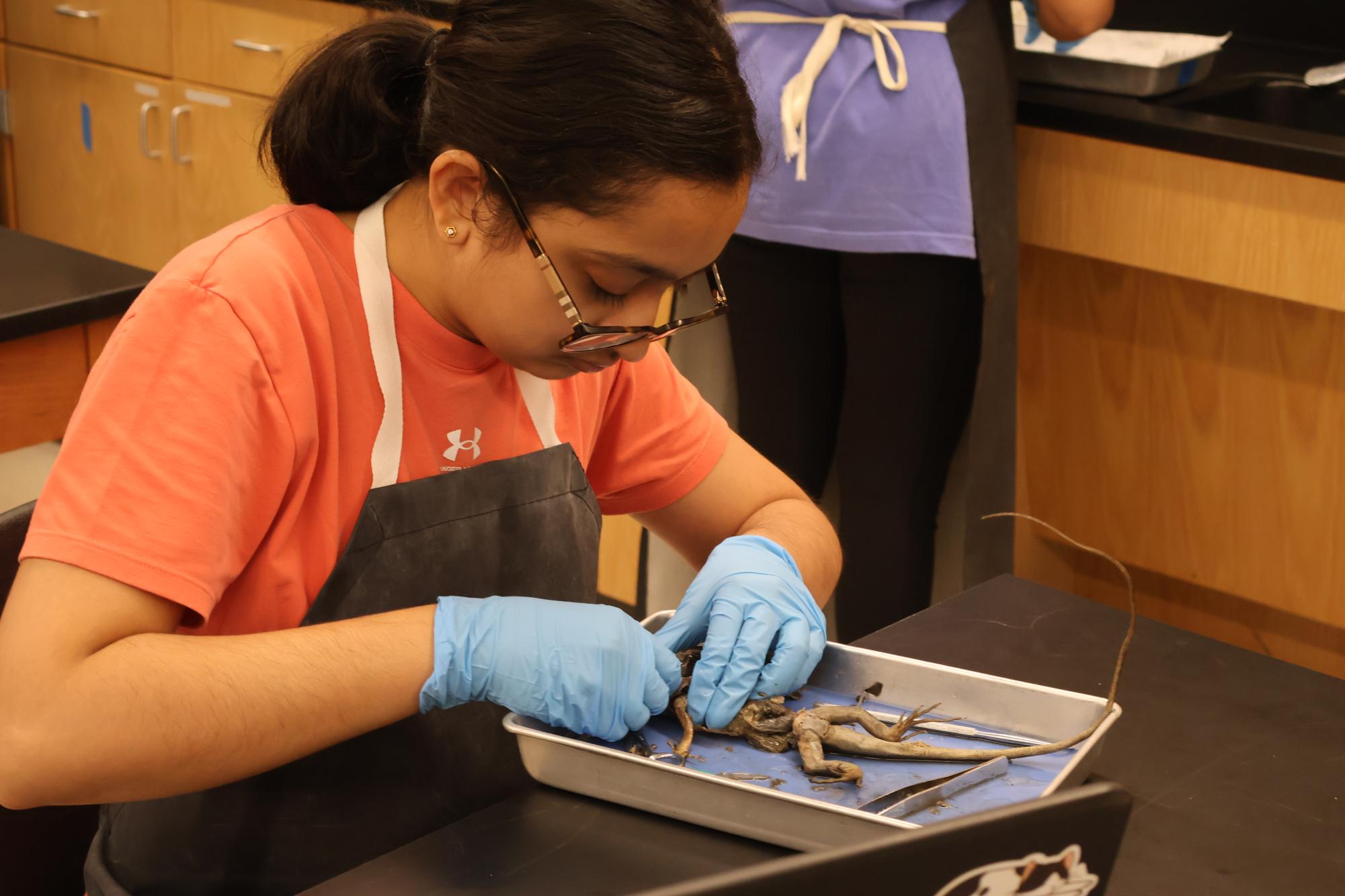
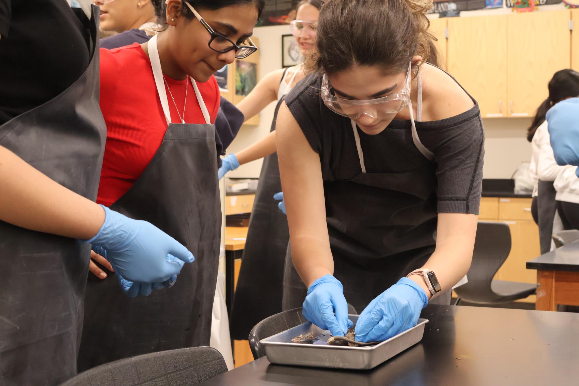
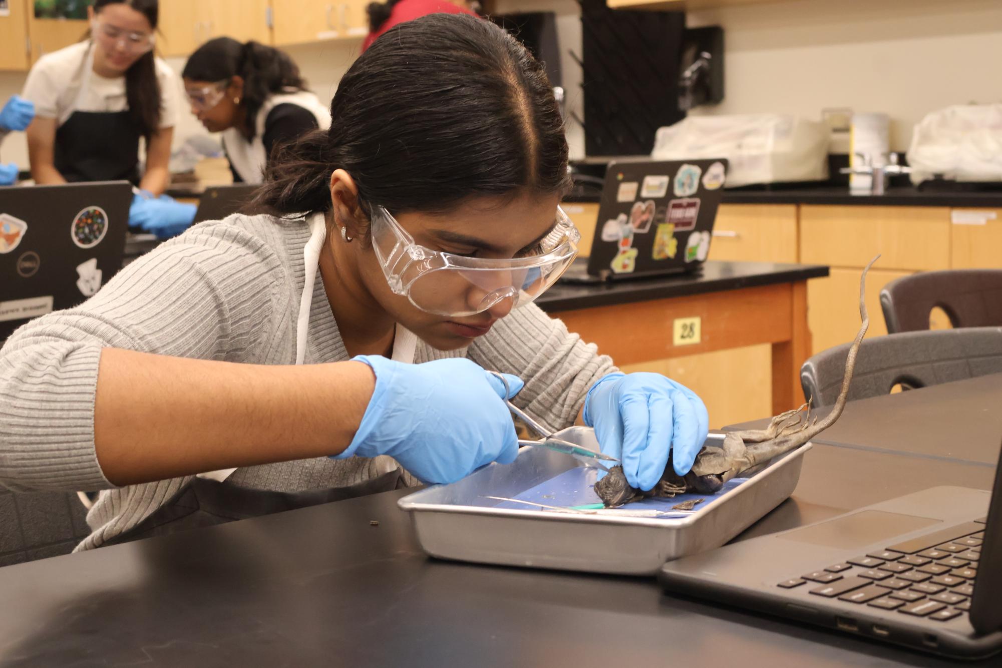
Afterward, the students skinned their iguanas, a time-consuming process that allowed them to remove the scale layer, revealing the organ systems underneath. The skinning process involved lifting the skin, making incisions, and cutting off the scale layer in large flaps using scissors.
“Peeling the skin from the reptile was a whole new experience, with the scaly texture, and there were these different parts like the ear,” Hasya Pamu ‘26 said. “The difficult parts were basically cutting it open without affecting those fragile organs within and getting through the tough skin, especially around the tail area, where I wanted [to see] the bone structure.”
Skinning the specimens made it easier for the participants to observe anatomical structures such as the dewlap, the multipurpose flap of skin beneath iguanas’ throats. They could also more easily see where the spine extended to support the dorsal crest, as well as the muscular differences between the front and back legs. Identifying all of the structures became a challenge, because they were all connected by one internal membrane.
“It was very difficult to actually see the organs inside, with a lot of different deflated sacs within,” Pamu said, “but it was really interesting to see this new organism. I don’t think we’ve ever done this before in our dissections.”
Anatomy & Dissection Club will meet again to dissect various specimens on Friday, April 25 at 8 a.m. in Mr. Scheiber’s room.






Pritt Changwatchai • Mar 31, 2025 at 7:56 pm
Sounds really interesting!